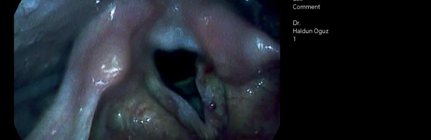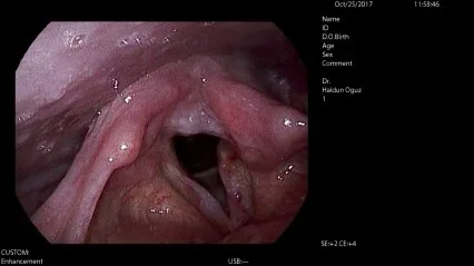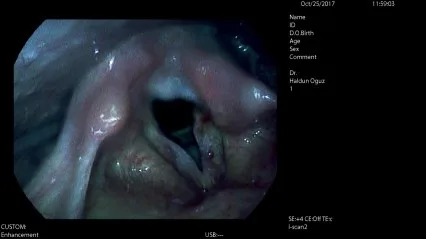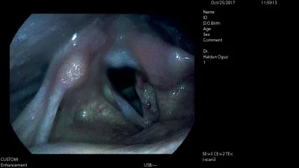

Early detection of vocal cord cancer (vocal fold cancer) requires accurate diagnosis and timely initiation of treatment. High-resolution endoscopic examination using digital contrast methods such as i-scan or narrow band imaging (NBI) can be useful in this regard.
How does I-scan contribute to diagnosis?
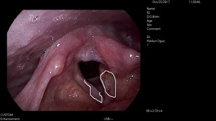
I-scan technology enables image quality enhancement and detailing by using surface enhancement (SE), contrast enhancement (CE) and tone enhancement (TE) to improve the distinction between healthy and pathological tissue. SE makes the edges of the tissues more prominent, allowing better definition of edges. CE provides better definition of deeper areas or regions with lower colour intensity. TE enhances the pathological area by changing the RGB colour content of each pixel in the image of the organ. During the examination, one can switch between different levels of these enhancement methods. In the images below, you can see endoscopic photographs of a patient with unilateral vocal cord paralysis and pathological lesions in both vocal cords.